Bacterial Information Transfer
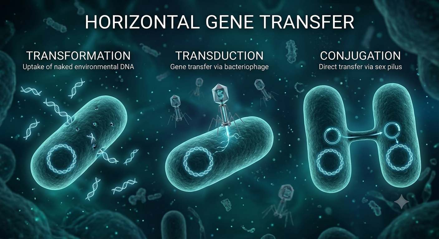
Bacterial information transfer revolves around how genetic code is maintained, expressed, and shared. Beyond standard central dogma processes (DNA replication, transcription, and translation), bacteria utilize Horizontal Gene Transfer (HGT) to share genetic material laterally between co-existing cells rather than passing it down vertically from parent to offspring.
Table 1. Bacterial Horizontal Gene Transfer (HGT) Mechanisms
Mechanism | Key Vector / Medium | Physical Requirement | Primary Biological Action |
|---|---|---|---|
Transformation | Naked environmental DNA | Cellular competence (membrane permeability) | Imports and integrates free extracellular DNA fragments released from lysed cells. |
Transduction | Bacteriophage (viral intermediate) | Viral infection (lytic or lysogenic cycle) | Packages host bacterial genes into viral capsids and injects them into new host cells. |
Conjugation | Conjugative plasmid (e.g., F- plasmid) | Direct physical contact via a sex pilus | Transfers a single-stranded plasmid copy across a cell bridge to a recipient cell. |
Transformation
Bacteria pick up free, naked DNA fragments directly from their extracellular environment. This occurs naturally when donor cells lyse (burst) and release their contents. For a cell to undergo transformation, it must enter a physiological state called competence, allowing it to transport large, negatively charged DNA molecules across its cell wall and membrane to integrate into its genome.
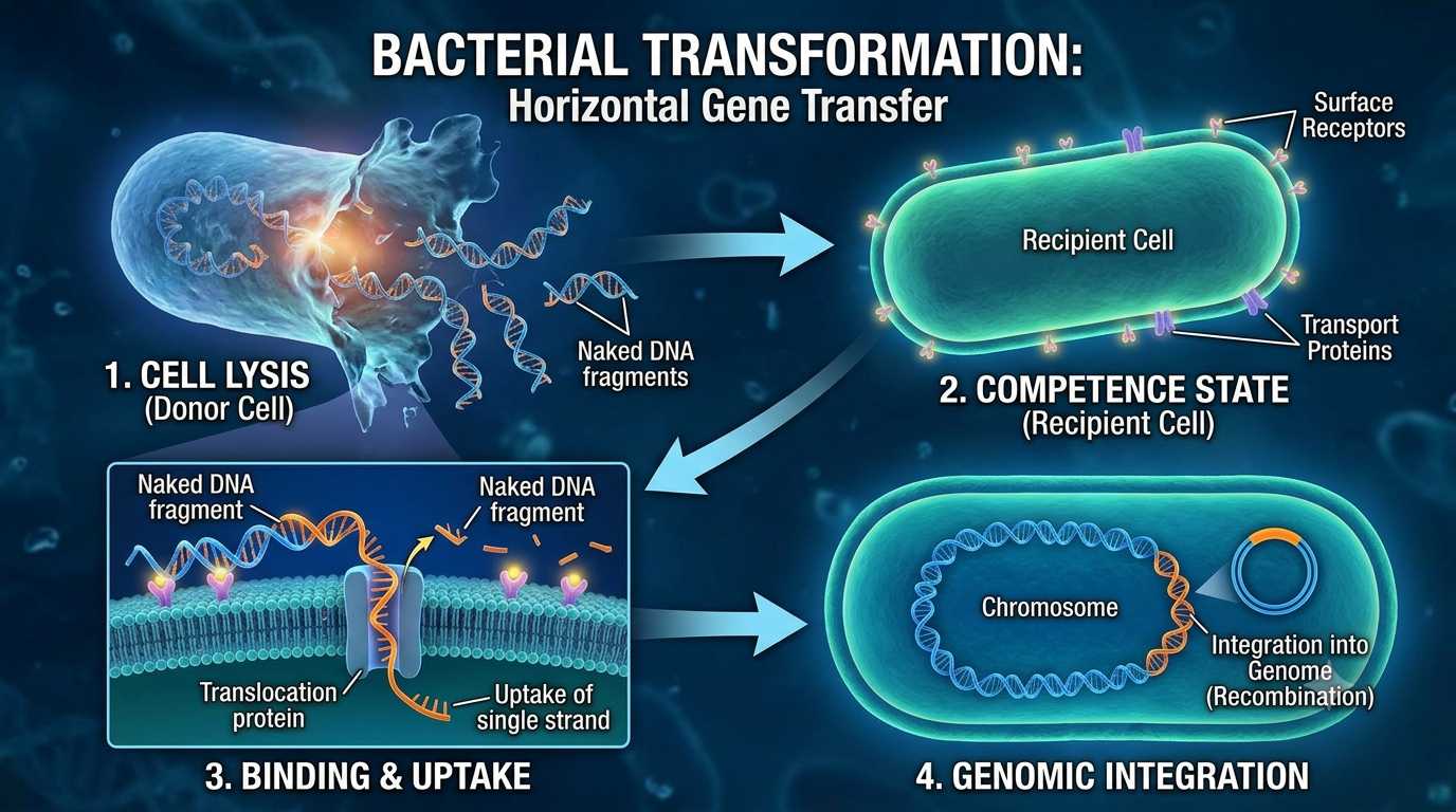 Picture 2. Horizontal Gene Transfer, Transformation
How the concept of Transformation can be visualised step-by-step:
Step 1: Cell Lysis (Donor Cell) A donor bacterial cell ruptures (lyses), releasing its genomic material and plasmid DNA as free, naked double-stranded DNA fragments into the surrounding extracellular environment.
Step 2: Competence State (Recipient Cell) A neighboring recipient cell enters a state of physiological competence, expressing specialized surface receptors and protein transport channels across its cell wall and plasma membrane.
Step 3: Binding & Uptake The free, negatively charged extracellular DNA binds to the surface receptors on the competent recipient cell. Membrane-bound translocation proteins pull a single strand of the DNA across the cell membrane into the cytoplasm while degrading the complementary strand.
Step 4: Genomic Integration Once inside the cytoplasm, the newly acquired single-stranded DNA interacts with recombination proteins (such as RecA) and integrates into the recipient bacterium’s circular chromosome or resides as a stable plasmid, permanently altering its genetic profile.
Transduction
This mechanism is mediated by bacteriophages (viruses that infect bacteria). During the lytic cycle, a phage infects a host, replicates, and accidentally packages host bacterial DNA instead of viral DNA into new phage heads. When these phages lyse the cell and infect a new host, they inject the previous host's bacterial DNA. In the lysogenic cycle, viral DNA integrates into the bacterial chromosome as a prophage; when it later exits the chromosome, it can carry adjacent host genes with it to the next recipient cell.
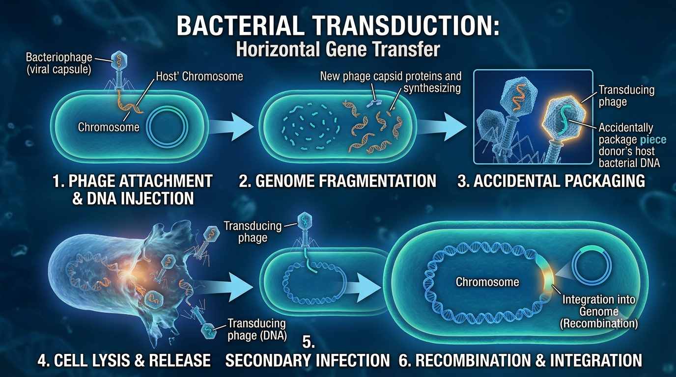 Picture 3 Horizontal Gene Transfer , Transduction
How the concept of Transduction can be visualised step-by-step:
Phage Attachment & DNA Injection: A bacteriophage binds to specific surface receptors on the host bacterium and injects its viral genetic material into the cell's cytoplasm, leaving the viral protein coat (capsid) outside.
Host Genome Fragmentation & Viral Replication: The injected viral DNA hijacks the host bacterium's metabolic machinery. It initiates the synthesis of phage proteins and directs host enzymes to degrade the host bacterial chromosome into small DNA fragments.
Accidental Packaging (Errors in Assembly): During the rapid assembly of new virus particles, a mistake occurs: instead of packaging newly replicated viral DNA, the phage capsid accidentally packages a fragment of the host bacterium's degraded DNA. This forms a transducing phage.
Cell Lysis & Release: The host bacterial cell lyses (bursts open) due to viral enzymes, releasing both normal bacteriophages and the newly formed transducing phages into the surrounding environment.
Secondary Infection: The transducing phage attaches to a new recipient bacterial cell and injects the original donor bacterium's DNA fragment into it. Because the phage carries bacterial DNA instead of viral genes, it does not cause a lytic infection or destroy the recipient cell.
Homologous Recombination & Integration: The injected donor bacterial DNA aligns with the recipient cell’s chromosome. Via recombination proteins (such as RecA), the donor DNA fragment integrates into the recipient’s genome, permanently updating its genetic traits (such as conferring antibiotic resistance).
Conjugation
Often referred to as bacterial mating, conjugation requires direct physical contact between a donor cell and a recipient cell. The donor cell possesses a conjugative plasmid (like the F-plasmid) that encodes the machinery to build a specialized protein bridge called a sex pilus. The pilus draws the two cells together, and a single-stranded copy of the plasmid is transferred across a conjugation channel into the recipient, which then synthesizes the complementary strand to complete the transfer.
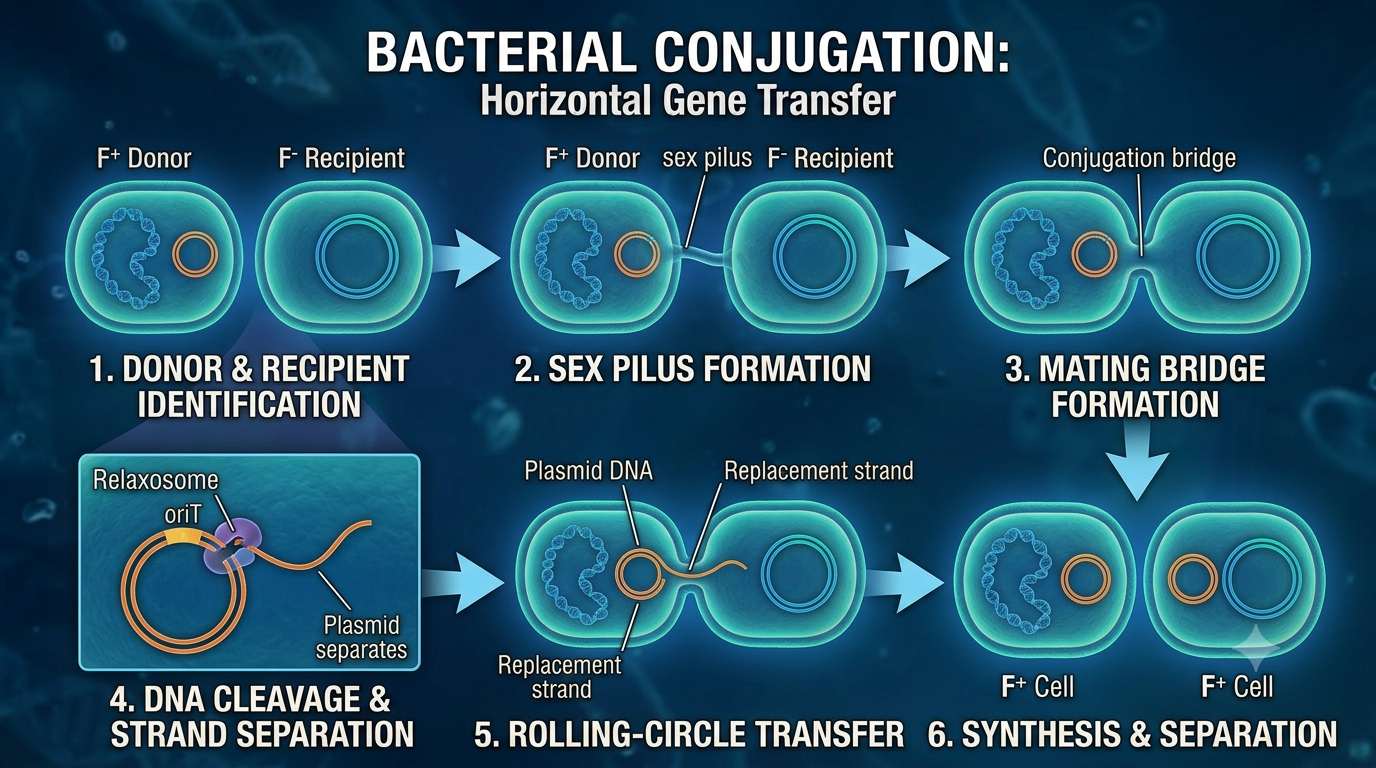 Picture 4. Horizontal Gene Transfer, Conjugation
How the concept of conjugation can be visualised step-by-step:
Donor and Recipient Identification: Two bacterial cells reside near each other: a donor cell (F+) containing a conjugative plasmid (such as the F-factor) and a recipient cell (F-) lacking the plasmid.
Sex Pilus Formation & Contact: The donor cell extends a specialized protein filament called a sex pilus (or conjugation pilus). The pilus physically attaches to specific surface receptors on the recipient cell membrane, anchoring the two cells together.
Mating Bridge Formation & Filament Retraction: The sex pilus depolymerizes (shortens), drawing the two bacterial cell walls into close proximity. A stable protein-based channel, known as the mating bridge or conjugation pore, forms across the cell membranes to connect both cytoplasms.
DNA Cleavage & Strand Separation: An enzyme complex (relaxosome) inside the donor cell recognizes a specific sequence on the plasmid called the origin of transfer (oriT). It makes a single-stranded cut (nick) in the plasmid DNA and unwinds one strand to prepare it for transport.
Single-Stranded Rolling Transfer: The relaxed, single-stranded DNA pass through the mating bridge into the recipient cell from the 5' end to the 3' end. Simultaneously, rolling-circle replication in the donor cell synthesizes a replacement strand so the donor retains a complete plasmid.
Complementary Strand Synthesis & Cell Separation: Inside the recipient cell, DNA polymerase synthesizes a complementary strand to convert the transferred single-stranded DNA back into double-stranded plasmid DNA. The mating bridge breaks, leaving both bacteria as fully functional donor cells (F+) capable of initiating conjugation with other cells.
Practical Implications of Horizontal Gene Transfer in Research and Bio-Engineering
Horizontal Gene Transfer (HGT) represents a fundamental paradigm in microbial genetics, governing the lateral exchange of genetic material across distinct bacterial populations. In the contemporary laboratory setting, the mechanics of HGT directly influence both biosafety protocols and advanced genetic manipulation strategies. Understanding these biological transfer pathways provides essential context for maintaining experimental integrity and optimizing gene delivery methodologies in bench-scale research.
Biosafety Implications and Sterile Technique
From an experimental safety perspective, the mechanisms of HGT elucidate the rapid dissemination of antimicrobial resistance determinants and pathogenic virulence factors within microbial communities. The persistence of extracellular, naked DNA in aqueous solutions highlights the continuous risk of transformation, while the stability of bacteriophages across physical surfaces facilitates unintended transduction events. Consequently, rigorous decontamination measures—including enzymatic degradation via DNases, high-pressure steam sterilization (autoclaving), and the targeted application of broad-spectrum chemical disinfectants—are essential to prevent cross-contamination and preserve the clonal integrity of research strains.